
|
|
|
| Disclaimer: | The contents of these pages are for information only. They do not serve as recipes for the reader to be used in any medical or other situation. Please do not hesitate to contact us if you feel that your or anybody else's intellectual property might be impinged upon. |
Heparin Coatings(by J.J.G. Bos) What a Heparin Coating Can DoDemonstration of Activity Applications Coating versus Injection Note concerning Friction Reduction What a Heparin Coating Can Do
Heparin is commonly used as antithrombogenic agent. Its main purpose is to prevent blood from clotting. It helps antithrombin III (AT3) to inhibit thrombin, an enzyme that induces blood coagulation. Heparin is known to interfere with other parts of the blood coagulation cascade as well.
return to top... Demonstration of Activity
The illustration below demonstrates the thrombin inhibition caused by heparin. Thrombin is generated in the blood as part of the coagulation cascade. Increased thrombin levels are usually associated with increased blood coagulation. Thrombin is inhibited by anti-thrombin III (AT3). Inhibition is greatly accelerated in the presence of heparin.
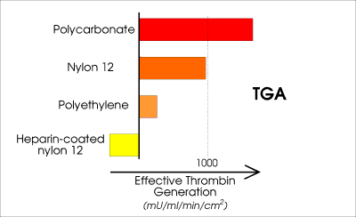 Thrombin inhibition still prevails over thrombin generation after extraction of this particular coating in phosphate-buffered saline (pH7.3) at body temperature for some 40 minutes. This is generally longer than a standard catheterization procedure lasts. Animal tests (porcine model) revealed at least 4 hours of sustained antithrombogenic action of this particular coating. The clinical test lasted this long - unfortunately no longer tests could be conducted with these samples, in order to determine the actual limit of the coating's operating window. return to top... Applications
Two barriers are commonly encountered when it comes to application of heparin coatings:
Cetry is specialized in breaking down both barriers by optimizing coating economy and assisting in generating the proper documentation for purpose of registration.
Heparinized endovascular catheters cause less generation of thrombi than the uncoated versions. This in turn reduces thrombi-related risks such as distal embolization. Unofficial data indicated a reduction from 7 out of 2000 downto 1 out of 2000 patients with major cerebral complications (i.e. blood clots blocking blood vessels feeding the brain) as caused by routine cardiovascular procedures. In other words: Induced near the heart, clots caused trouble in the brain. Not all devices involved in these procedures - such as the guide wires and the catheter sheath introducers - were heparinized, just the diagnostic catheters. And still the effect was very significant! Typical base materials involved during these procedures were: nylon, polyurethane (linear chains), polyethylene. Typical exposure times: 10 - 20 minutes. Sets for blood transfusion are generally made with use of PVC tubing. By coating the inside of the tubing (or the entire set) with heparin, it becomes possible to significantly reduce blood coagulation. It may also be profitable to coat the outer surface at the parts that are inserted into the veins. Intraocular lenses with heparin coating are commercially available nowadays. The heparin helps reduce inflammatory response. Typical base material: polycarbonate. It may be advantageous to coat both the inner and outer surface of tubing (and valve) of hydrocephalus drains, in order to reduce inflammatory response.
I have some photographs included on the web page about
heparin in tubing. The pictures show heparin coatings applied to nylon, silicone, polyethylene and PVC, but application to many other materials is well possible such as PU and FEP. 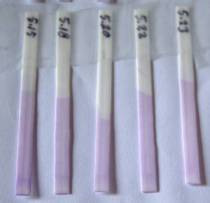
return to top... Coating versus Injection
Patients are usually injected with heparin as part of the endovascular procedure. This is done in order to increase blood coagulation times, so that the chance of complications due to (ongoing) clotting is significantly reduced. This helps, because now there is more time for fresh blood to dilute the activated blood that comes from the area of intervention. There is thus more time and chance for the blood to restore its original biochemical balance.
A heparin coating with activity of 0.1 IU per square cm and an impact range into the blood of e.g. 5 microns shows a very localized impact, equivalent to 200,000 IU per liter blood. Safe systemic injections, however, involve doses of 5,000 - 10,000 IU heparin for adult persons. This relates to some 1,000 - 2,000 IU heparin per liter blood. For blood localized within the 5-micron zone around an uncoated medical device, this is a hundred times less than that of the heparin-coated device. It illustrates how the coating works as a barrier. The closer to the device, the greater the effect. Do note that the blood outside the action range of the heparin coating is not affected by clotting, due to absence of contact activation. return to top... Note concerning Friction Reduction
Heparin is a hydrophilic molecule, i.e. it attracts water, but most heparin coatings are not slippery as is the case for some hydrogels. The coefficient of friction of heparin in water is close to that of for instance nylon or (linear) polyurethane, thus in now way close to that of polytetrafluoroethylene (PTFE-teflon). However, the coefficient of friction experienced between uncoated nylon surfaces (or PE, PU, etc.) may increase when in contact with living blood due to thrombus formation, whereas friction between heparin-coated surfaces tends to remain constant. Cetry has successfully developed a low-friction heparin coating, with a coefficient of friction comparable to that of PTFE and HDPE. Information regarding Cetry's own low-friction heparin coating can be found HERE.
return to top... |